Monoclonal Antibody Production
 | During the production of monoclonal antibodies using hybridoma technology, a mouse is immunized with a selected antigen. Collections of immunized spleen cells from the mouse are fused together with myeloma cancer cells to produce hybridoma cells. Because spleen cells are fused together with cancer cells, hybridoma cells are essentially immortal and can be used in production as many times as needed. When a large quantity of hybridoma cells have grown, they are diluted down, and their antibodies are screened for a reaction against the selected antigen. The strongest cell clone is isolated and grown in a cell culture. |
Hybridoma cells will mulitply and produce large quantities of identical, antibody-forming immune cells. At this point, several filters are used to separate the specific antibodies from the remaining unwanted cell debris. Once isolated, these antibodies are grown into colonies, and a single population of mAb can be extracted for the use of medicine.
Since mice, now even rabbits, are used in this process, they are put in a harmful position during testing. Because of this risk, monoclonal antibody production is looked at as unethical. Cell cultures are used as often as possible to limit any possible harm.
Antibody Stucture and Formation
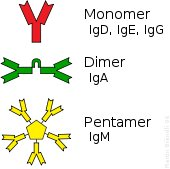
Antibodies, also known as immunoglobulins (Ig), are proteins produced by the body's B-cells and T-cells that identify, attach, and bring down antigens of foreign pathogens. The basic structure of the antibody includes two large heavy chains and two small light chains to form three figures: monomers (one unit), dimers (two units) and pentamers (five units). Each unit contains two antigen-binding sites, where they then bind to specific binding sites on the antigen called epitopes. Because of the organisms' genes, individual antibodies have diverse antigen-binding sites, thus allowing various antibodies to exist and to attack the various antigens as well. Antigen-binding sites are able to quickly recognize and bond specifically to an epitome among the millions of other antigens in the body. Once an antibody is attached, it signals other immune defense cells to attack. The different types antibodies are called isotopes and vary between species of mammals.
| 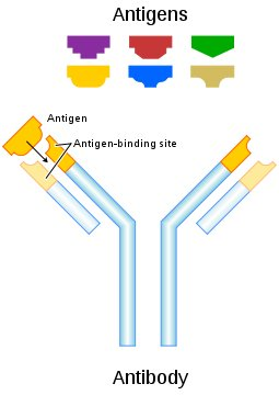 This picture represents a single unit of an antibody that bonds with a specific antigen. The different colored shapes represent the various shapes and sizes of antigens. |